Foot Anatomy — Tissue & Bone & Ligaments & Joint
The foot is the only tissue in the human body that is in continuous contact with the ground, so the foot needs to have enough cushioning force to absorb the weight to adapt to the space force of the ground. At the same time, the foot needs tough power to complete the driving force of fast walking or running.
Therefore, the evolution and function of the foot is the focus of research in the current rehabilitation exercise. The bone structure of the foot has many similarities with the bone structure of the hand. The radius of the forearm and the tibia of the leg are both important to form small bones. The joints of the wrist and the tarsal bones of the foot are like steel beans in the bearing, which play a key role in the linkage of the joint joints. The metatarsals of the wrist and the metatarsals of the foot are like the metatarsals connected to their respective distal ends. There is a big similarity with the phalanx.
Foot soft tissue
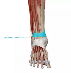
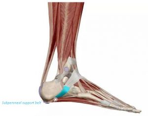
The support belt of the ankle joint is a fibrous band of tissue located in the ankle joint. It is formed by thickened deep fascia. The main function is to fix the tendon and prevent the single bundle from stretching out.
The upper extensor support belt: Located in the front and upper part of the tibial calf joint, it wraps around the anterior tibial anterior tendon, extensor pollicis longus tendon, extensor digitorum longus tendon, and third peroneal tendon. The support straps on the extensors are wider and run around the ankles.
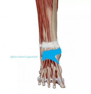
Inferior extensor support belt: in a y shape from the bifurcation of the basal bone around the ankle to the medial malleolus and navicular bone, located in front of the tibiotalar joint, also known as the “calf cruciate ligament” mainly wraps around the extensor digitorum longus and the third The three peroneus muscles.
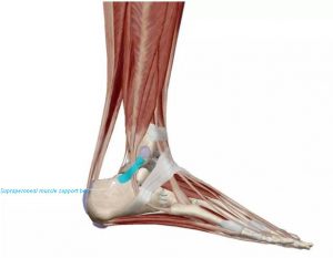
Supraperoneal muscle support belt: Located on the outside of the calf and root bone, it is also an extension of the deep fascia. Damage to this support belt can make our tendons unstable.
Subperoneal support belt: It continues with the lower support belt for extensor muscles, combines with the gastrocnemius tendon and the peroneus brevis tendon, and enters the foot.
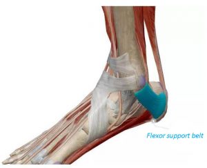
Flexor support belt: in the upper and front of the medial malleolus of the foot, it has a rectangular shape and wraps the flexor pollicis longus, flexor digitorum longus, and posterior tibial muscle. The bone fibrous tube formed by the flexor support belt, the tibial medial malleolus, and the calcaneus is the ankle tube.
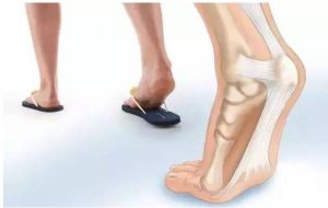
Plantar fascia
The plantar fascia is a slender fiber bundle located on the plantar surface of the plantar. It extends from the heel to the toe. It has a large span and is composed of dense collagen fibers. They are basically arranged vertically and attached to the flexor digitorum longus and Above the flexor digitorum brevis, it extends on both sides to the first and fifth phalanx. The central part is the thickest and toughest. It starts from the inner side of the calcaneal tubercle. The inner part of the plantar fascia is more It is thin, covering the abductor pollicis, and extending proximally to the flexor support belt.
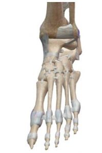
Skeletal anatomy: The foot can be roughly divided into three joints composed of a group of bones and one or more joints. The hindfoot is composed of the talus, calcaneus, and subtalar joint; the midfoot is composed of the remaining tarsal bones; including the transverse tarsal joint and the smaller distal intertarsal joint; the forefoot is composed of the metatarsal bones and phalanges, including the tarsal and metatarsal joints and their distal ends The foot consists of 26 intricate ligaments, plantar aponeurosis, tendons, muscles, and other soft tissues to form a stable joint structure and arch structure.
Bone and Ligaments:
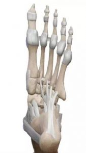
7 tarsal bones: talus, calcaneus, navicular bone, medial, middle, and lateral cuneiform bones, cuboid bones
5 metatarsals
14 phalanges: proximal, middle, and distal
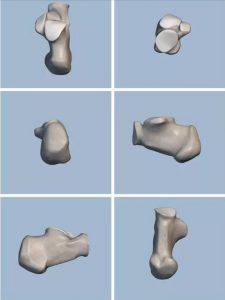
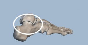
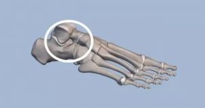
Calcaneus:
The largest tarsal bone, the front 2/3 is called the calcaneal body, and the back of the body is short with thick, rough calcaneal nodules. The shape of the calcaneus has 6 sides:
Above: through 3 articular surfaces (front, middle, and back) and the talus related nodes;
Front: the section related to the cuboid bone;
Behind: the section related to the stop point of the Achilles tendon;
Medial surface: There is a bony process called the intercept process, and the supporting part of the talus head is the attachment point of many ligaments at the same time;
Lateral surface: bony bulge, called fibular trochlea, which separates the fibular long and short tendons;
Below: The main weight-bearing surface of the hindfoot is the attachment point of many internal muscles and ligaments of the plantar fascia.
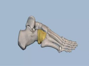
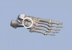
Navicular bone:
Back: connect with the head of the talus;
Front: connected with the 1st and 3rd cuneiforms;
Inside side: there is a round bulge facing downward, called the scaphoid trochanter;
Cuboid bone:
Back: Connect the calcaneus
Front: Connect the 4th and 5th metatarsals
Bottom: There is a round bulge called cuboid tuberosity. In front of it is the lateral groove through which the peroneus longus muscle membrane passes. To
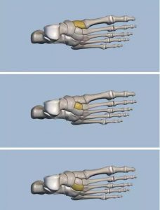
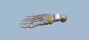
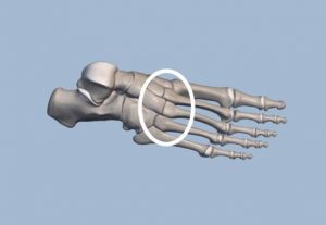
Cuneiform
The first to third cuneiforms are arranged from the inside to the outside, and gradually decrease in sequence, with the wide side of the second and third cuneiform facing upwards, and the narrow side of the first cuneiform facing upwards, and the mutual fit is stable.
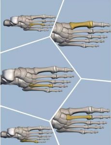
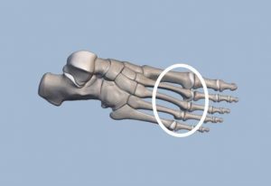
Metatarsals
It is a short tubular long bone, 5 pieces in total (from the inside to the 5th metatarsals in turn)
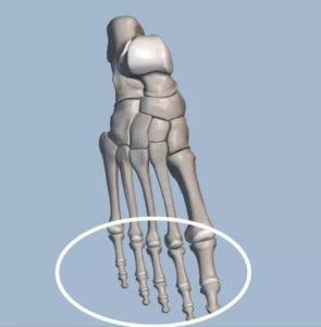
Phalanges
There are 14 in total, except for the two toes, and all the other toes are three. The phalanges are divided into bottom, body, and pulley.
ligament:
Talar Ligament:
Including the anterior, posterior, and internal talar ligaments, the lateral talar ligament bundles, the oblique talar heel bundles, and the interstellar calcaneal ligaments.
The main function is to stabilize the talar-heel joint and limit excessive varus and valgus of the joint

Dorsal ligament:
Including the dorsal ligament of the talus scaphoid, the divergent ligament, the dorsal ligament of the calcaneus cuboid, the dorsal ligament of the cuneate cuneiform, and the dorsal ligament of the cricoid scaphoid.
The main function is to stabilize the structure of the intertarsal joints such as the talar-calcanean navicular joint and the calcaneocuboid joint.
Plantar ligament:
Including the long plantar ligament, the short plantar ligament, the cuneiform plantar ligament, the cuneiform plantar ligament, and the navicular plantar ligament (spring ligament).
The main function is to maintain the normal shape of the arch of the foot.
joint
There are more than 30 joints in the foot, mainly including intertarsal joints (talar-heel joint, talar-navicular joint, calcaneocuboid joint), tarsometatarsus joint, inter-metatarsal joint, metatarsophalangeal joint, and toe Between joints. To
(1) Talar-heel joint (subtalar joint)
The anterior, middle and posterior articular surfaces above the calcaneus form the talar-heel joint with the talus.
The anterior subtalar joint is composed of a concave anterior and middle articular surface of the calcaneus and a protruding anterior and middle articular surface of the talus; the posterior subtalar joint is composed of a convex posterior articular surface of the calcaneus and a concave subtalar articular surface. This cross change of concave or convex articular surface enables complex torsion activities.
(2) Talar and navicular joint
The articular head is the head of the talus, and the articular socket is composed of the articular surface of the talus behind the navicular bone and the front and middle articular surfaces of the upper calcaneus.
From the morphological point of view, the talar-navicular joint and talar-calcaneal joint are two independent joints. But from a functional point of view, the two are joint joints that move along a common axis of motion. The talar and navicular joint also has the function of foot varus and valgus.
(3) Transverse tarsal joint
Including the talar navicular joint and the calcaneal-cubic joint: although the two have their own independent activities, they need to complete their functional activities together.
The joint line of the transverse tarsal joint is curved in an “S” shape across the middle of the tarsal bone group, with the inner part protruding forward and the outer part protruding backward.
The main function is to better coordinate with the adjacent joints, especially the talar-heel joints, to complete the pronation and supination movements of the foot.
(4) Tarsal and metatarsal joints
A planar joint is composed of the base of the first to fifth metatarsals, cuneiform bones, and cuboid bones.
The tarsal and metatarsal joints can do slight sliding and flexion and extension movements, and participate in slight adduction and abduction movements.
(5) Metatarsophalangeal joints
The small head of each metatarsal bone and the middle bottom of the proximal phalanx of each toe is composed of five in total.
Roughly similar to the corresponding joints of the hand, but with a more stable structure and flexible movement.
(6) Interphalangeal joint
It is located between two consecutive phalanges, it is composed of the phalanx and the bottom of the distal phalanx.
To flex the joints, you can do flexion and extension exercises. The movement of the proximal interphalangeal joint is greater than the movement of the distal interphalangeal joint.